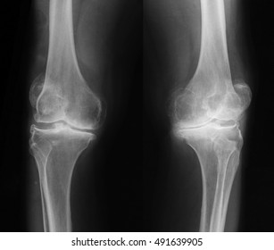
Bilateral X Ray Knee


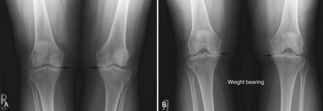
Knee Ap Weight Bearing View Radiology Reference Article

Knee Ap Weight Bearing View Radiology Reference Article

Ortho Dx Bilateral Knee Pain Clinical Advisor

Ortho Dx Bilateral Knee Pain Clinical Advisor

Bilateral Ap X Ray Of The Knees The Aberrant Position Of The

Bilateral Ap X Ray Of The Knees The Aberrant Position Of The

Teenage Girl S Knees Bilateral X Rays Stock Image C040 3070

Teenage Girl S Knees Bilateral X Rays Stock Image C040 3070

Bilateral Total Knee Replacement X Ray Stock Image C004 1483

Bilateral Total Knee Replacement X Ray Stock Image C004 1483

Bilateral Knee X Ray Showing Patella Alta Download Scientific

Bilateral Knee X Ray Showing Patella Alta Download Scientific

Bilateral Knee X Ray Showing Patella Alta Download Scientific

What can be seen on a knee x ray.

Bilateral x ray knee. You should instead list the appropriate radiology code twice on the claim form. 73564 x ray exam knee 4 or more 73565 x ray exam of knees procedure code modifier description 2015 payment rate 2016 payment rate percent change in payment rate 73562 x ray exam of knee 3 34 50 35 83 3 9 73562 26 x ray exam of knee 3 10 06 9 67 3 9 73562 tc x ray exam of knee 3 24 43 26 15 7 0. Bilateral x rays sometimes your doctor may want to have an x ray done on both knees. Medicare doesn t subject 73562 radiologic examination knee.

73564 x ray knee 4 views 73565 x ray bilateral knees standing 73590 x ray tibia fibula 2 views 73600 x ray ankle 2 views 73610 x ray ankle 3 views 73620 x ray foot two views 73630 x ray foot 3 views 73650 x ray heel 2 views 73660 x ray toe 2 or more views 71100 xray ribs unilateral. When caught early the condition may be managed so that you can stop the degenerative wear and tear. This x ray shows a healthy joint with nice sharp well defined edges at the joint margins. The patella or kneecap is seen sitting in front and to the left of the femur.

In a good lateral image the medial and lateral femoral condyles project over each other and the patellofemoral joint is projected free. This is called a bilateral x ray and is especially common if your doctor is checking for signs of arthritis. Three views to bilateral procedure payment rules which pay certain bilateral procedures at 150 percent. This view clearly shows the four knee bones.

The x rays pass through the knee joint from medial to lateral fig. Technique for lateral image of the right knee mediolateral projection. The lateral knee view is an orthogonal view of the ap view of the knee the projection requires the patient to roll onto the side of their knee hence it is not an appropriate projection in trauma in all suspected traumatic injuries of the knee the horizontal beam lateral method should be utilized. For instance if your physician takes three views of each knee you should report.

Femur tibia fibula and patella.
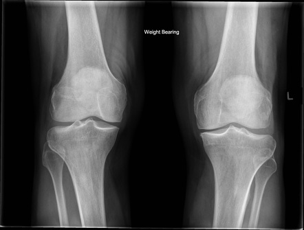
Bilateral Osteoarthritis Knee Joint Xray Image Stock Photo Edit

Bilateral Osteoarthritis Knee Joint Xray Image Stock Photo Edit

Radiographic Positioning Examples Of The Leg And Knee Ce4rt

Radiographic Positioning Examples Of The Leg And Knee Ce4rt

Bilateral Knees 10 13 16 Copy Dynamic Yoga Therapeutics

Bilateral Knees 10 13 16 Copy Dynamic Yoga Therapeutics

Patient With Bilateral Knee Osteoarthritis Discusses Stem Cell

Patient With Bilateral Knee Osteoarthritis Discusses Stem Cell

Lateral X Rays Of Bilateral Knees Showing Bilateral High Riding

Lateral X Rays Of Bilateral Knees Showing Bilateral High Riding

Science Source Bilateral Knee Osteoarthritis X Ray

Science Source Bilateral Knee Osteoarthritis X Ray

Normal Knees X Ray Stock Image F003 3510 Science Photo Library

Normal Knees X Ray Stock Image F003 3510 Science Photo Library

Prolotherapy Saved Me From Bilateral Knee Replacements Journal

Prolotherapy Saved Me From Bilateral Knee Replacements Journal

Knee Series Radiology Reference Article Radiopaedia Org

Knee Series Radiology Reference Article Radiopaedia Org

Knee X Rays And Detecting Abnormalities

Knee X Rays And Detecting Abnormalities

Knee Imaging Techniques And Normal Anatomy Musculoskeletal Key

Knee Imaging Techniques And Normal Anatomy Musculoskeletal Key

Figure 1 From Left Knee Pain And Bilateral Knee Swelling In An

Figure 1 From Left Knee Pain And Bilateral Knee Swelling In An

Simultaneous Bilateral Knee Varus Stress Radiographic Technique

Simultaneous Bilateral Knee Varus Stress Radiographic Technique

Ortho Dx Popping And Pain In An Adolescent S Knee Clinical

Ortho Dx Popping And Pain In An Adolescent S Knee Clinical

Source : pinterest.com