X Ray Knee Bilateral Ap Standing

Code 73656 can be most challenging says jandroep.
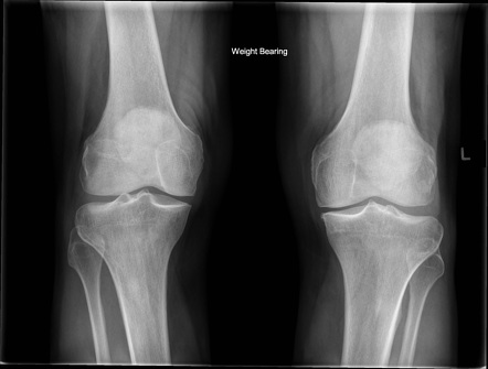
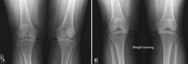
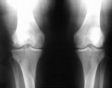
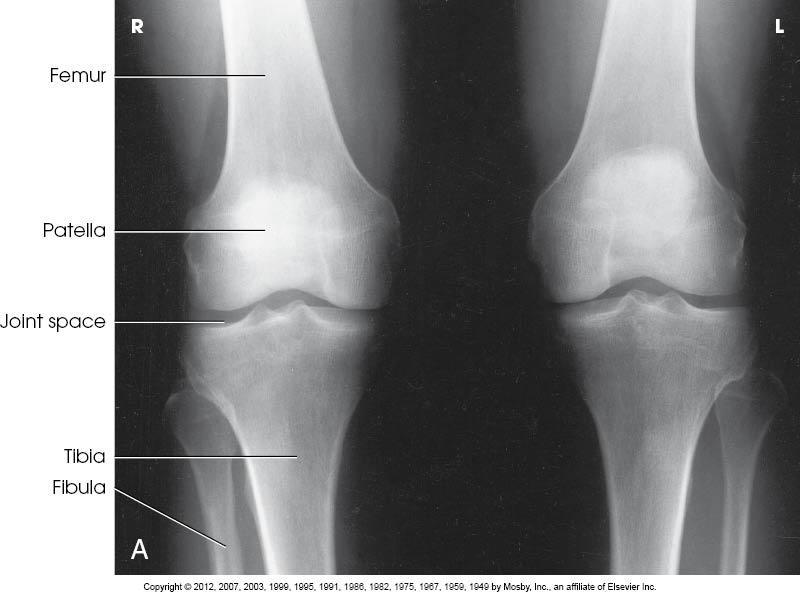
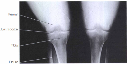
X ray knee bilateral ap standing. 73550 x ray femur 2 views 73560 x ray knee 1 2 views 73562 x ray knee 3 views 73564 x ray knee 4 views 73565 x ray bilateral knees standing 73590 x ray tibia fibula 2 views 73600 x ray ankle 2 views 73610 x ray ankle 3 views 73620 x ray foot two views 73630 x ray foot 3 views 73650 x ray heel 2 views 73660 x ray toe 2 or more views 71100. An alternative to the supine position is the standing ap image. You may read that your radiologist obtained a standing ap view x ray of the knee in addition to the oblique and lateral views you do not report code 73565. X rays taken while standing up can show the alignment of the knee joint and whether or not there is an abnormality in the alignment of the bone.
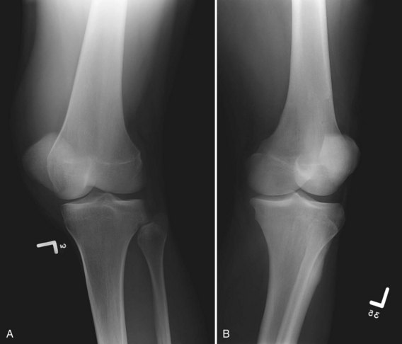
Patient position patient is supine on the table with the knee and ankle joint in contact with the table leg is extend. The front to back or anterior posterior knee image can be made in both supine and standing positions fig. 2 malalignment can lead to excessive forces on parts of the joint and accelerate arthritic changes. The knee ap weight bearing view is a specialized projection to assess the knee joint distal femur proximal tibia and fibula and the patella.
The knee ap view is a standard projection to assess the knee joint distal femur proximal tibia and fibula and the patella. The long common names have been created via a table driven algorithmic process. Oa is a painful degenerative condition that can reduce your mobility and make daily tasks difficult to manage. You instead count the standing ap view as a third view and you report code 73562.
The 45 flexed pa standing view of the knee is a much more sensitive x ray showing early degenerative disease in the position of function. In supine position the x rays pass through the knee from anterior to posterior ap image. Xr knee bilateral ap and lateral w standing this field contains the loinc term in a more readable format than the fully specified name.
